The mechanism of drug release from complex dosage forms, such as multivesicular liposomes (MVLs), is complex
and oftentimes sensitive to the release environment. This challenges the design and development of an appropriate
in vitro release test (IVRT) method. In this study, a commercial bupivacaine MVL product was selected as a
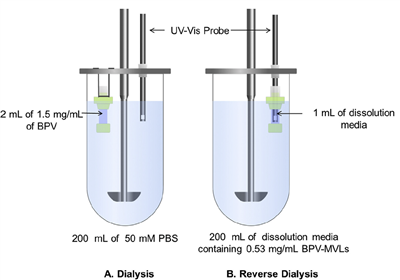
model product and an IVRT method was developed using a modified USP 2 apparatus in conjunction with
reverse-dialysis membranes. This setup allowed the use of in situ UV–Vis probes to continuously monitor the drug
concentration during release. In comparison to the traditional sample-and-separate methods, the new method
allowed for better control of the release conditions allowing for study of the drug release mechanism.
Bupivacaine (BPV) MVLs exhibited distinct tri-phasic release characteristics comprising of an initial burst release,
lag phase and a secondary release. Temperature, pH, agitation speed and release media composition were
observed to impact the mechanism and rate of BPV release from MVLs. The size and morphology of the MVLs as
well as their inner vesicle compartments were analyzed using cryogenic-scanning electron microscopy (cryo-
SEM), confocal laser scanning microscopy and laser diffraction, where the mean diameters of the MVLs and their
inner “polyhedral” vesicles were found to be 23.6 ± 11.5 μm and 1.52 ± 0.44 μm, respectively. Cryo-SEM
results further showed a decrease in particle size and loss of internal “polyhedral” structure of the MVLs over the
duration of release, indicating erosion and rearrangement of the lipid layers. Based on these results a potential
MVL drug release mechanism was proposed, which may assist with the future development of more biorelevant
IVRT method for similar formulations.
 4008709108
 info@pion.com.cn